MEYER DENTAL CBCT
3D PRO
Ultimate Sharp Vision, Powered
by Advanced Image Processing Technology
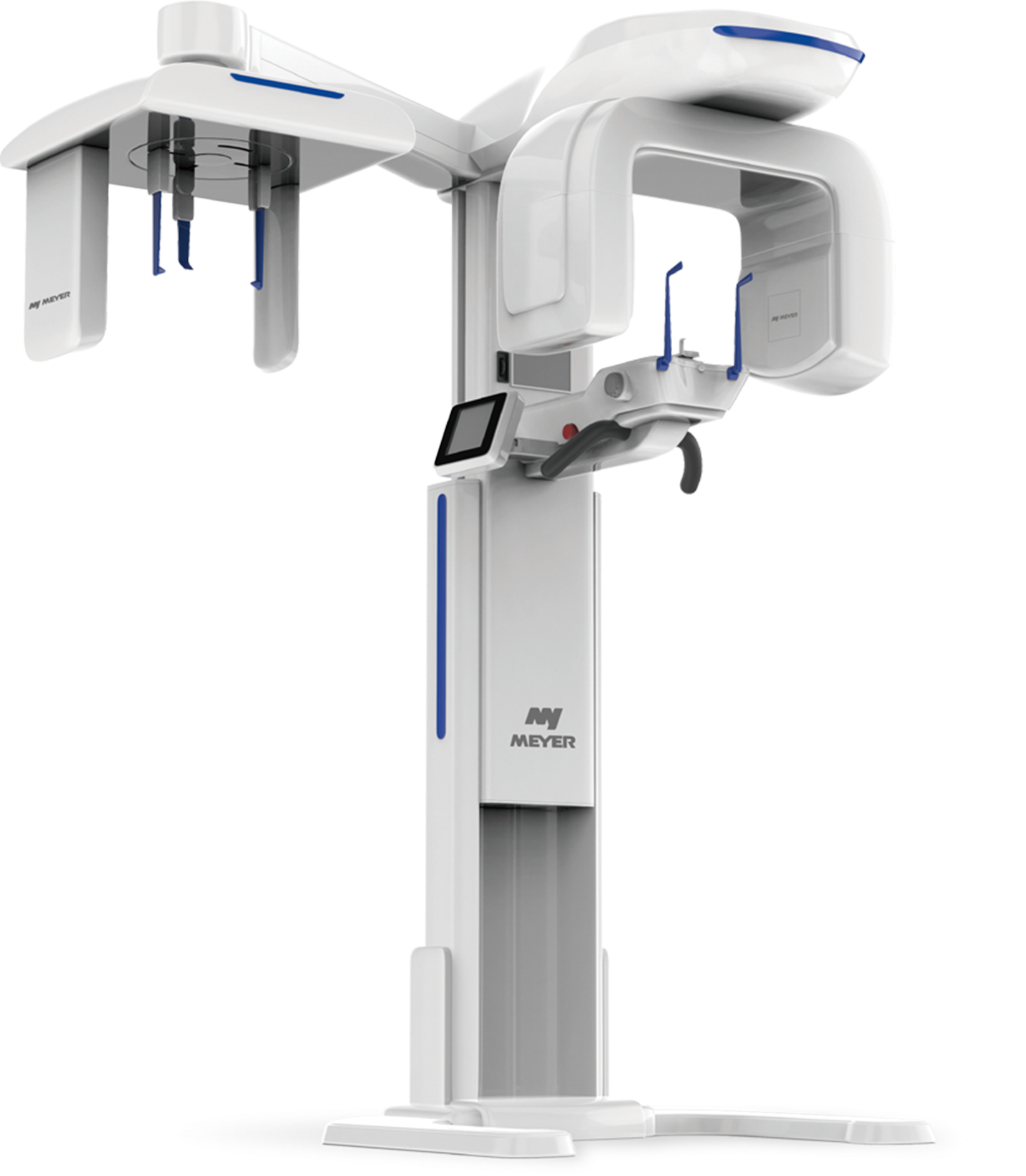
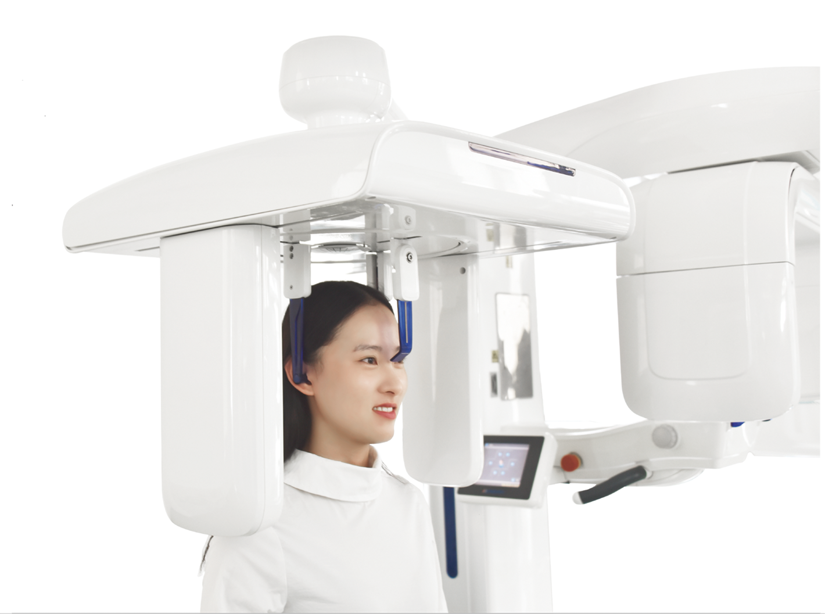
The Meyer extraoral CBCT equipment collects full oral data in one scan and reconstructs all aspects of high-resolution images as needed for accurate clinical diagnostics. The resulting 3D images and analytical data provide essential basis for dental filling, implant and orthodontics.
Overview
Highlights and Advantages
- 5-in-1 including CBCT, Pano, Ceph, Part CT and Model Scanning
- Robust processing algorithms to generate high image quality and accuracy
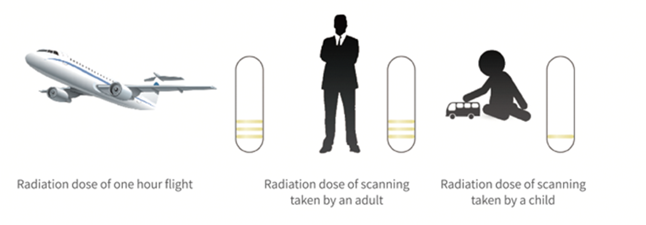
- Fast & precise with minimum-dose scanning. Child-safety mode available.
- Instant 3D image reconstruction post scanning through the new generation imaging technology
- Touchscreen for convenient operation
- Three-point patient positioning for stability and simplicity
Multiple FOV sizes
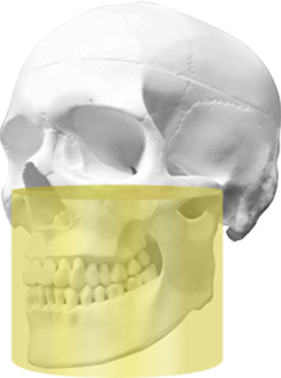
FOV12x10cm
- Covers from lower jawbone to maxillary sinus, and airway. - Suitable for general and local diagnostics. and preoperative evaluation of dental implant.
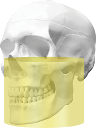
FOV15x 10.5cm
- Covers from lower jawbone to maxillary sinus, TMJ and airway
- Suitable for complete dentition scan,TMJ examination and preoperative evaluation of dental implant.
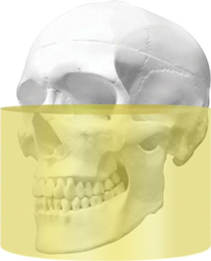
FOV17x11cm
- Covers from lower jawbone to maxillary sinus, TMJ and airway.
- Suitable for general and high
-resolution local diagnostics, TMJ examination and preoperative evaluation of dental implant.
Functions
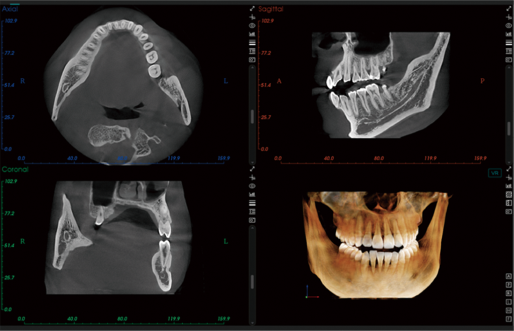
CBCT
Clear 3D anatomical display of maxillofacial area, general applied in various dental practice. Accurate 3D images by intelligent imaging technology, observation from any angle of view, accuratemeasurement of distance, surface area, volume and contour outline.
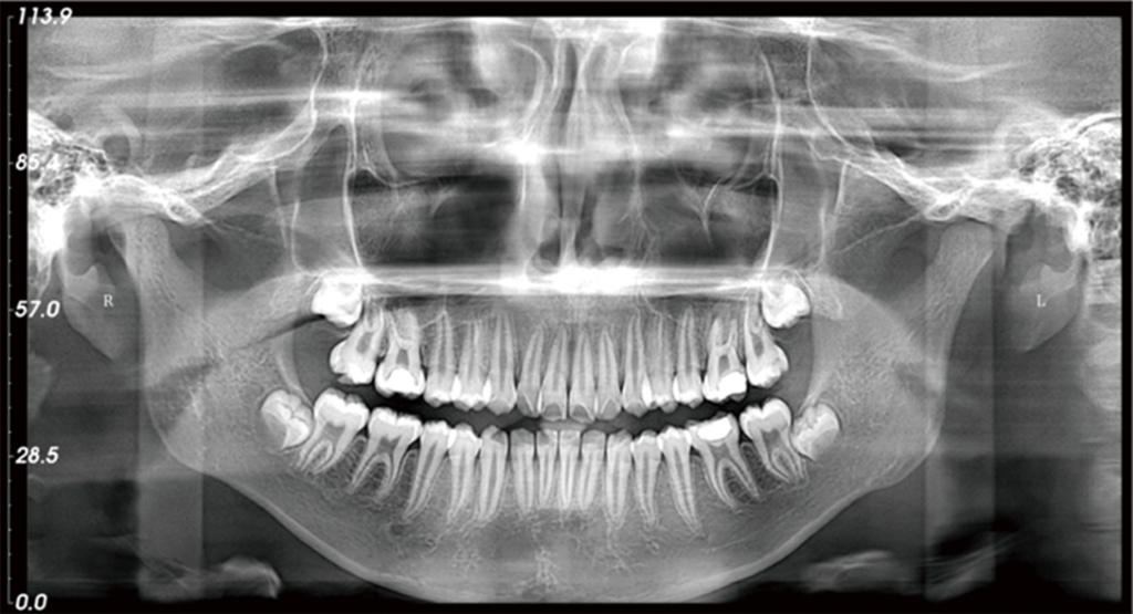
Pano
Optimized tomographic motion and structural rotation, allows for confident diagnosis of both mandible and maxilla areas.
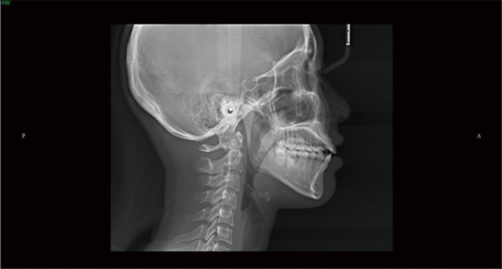
Ceph
Dual-level alignment of a single X-ray source to produce HD cephalometric images with low dose radiation.
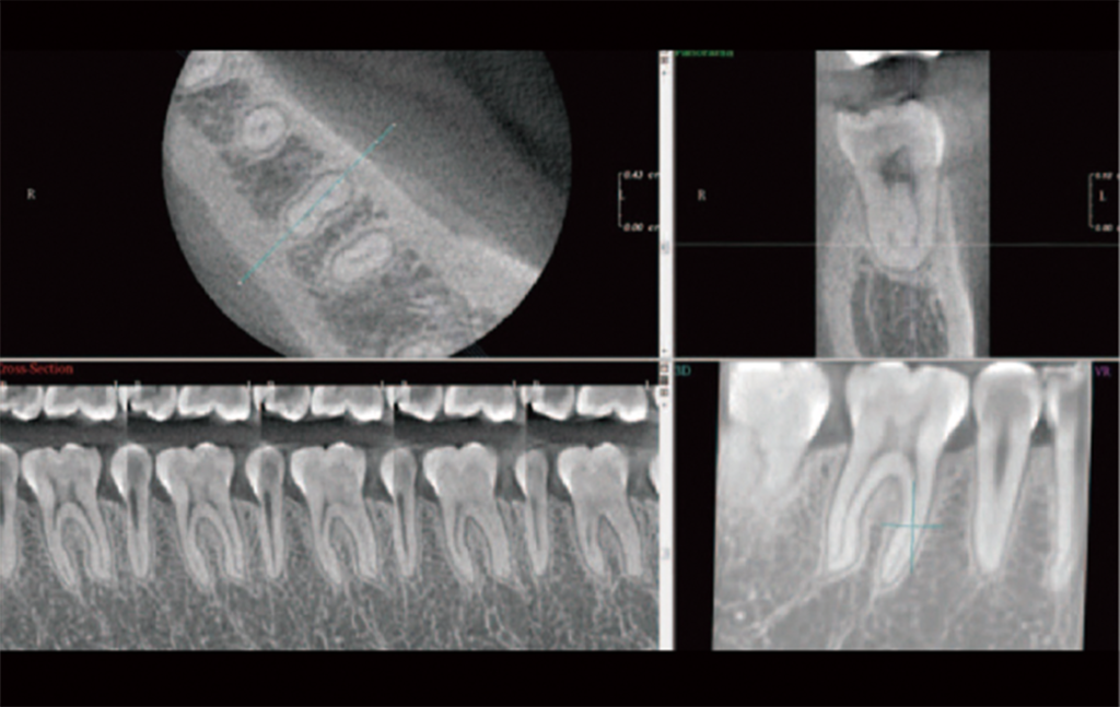
Partial CT
Under the partial CT mode, the image voxel can reach 40 80μm. It also reduces the DAP by 40%, which leads to lower radiation dose.
Model scanning
Just place the plaster cast or mold in the scanning area to scan. The data will be transferred to the software for subsequent design and clinical application.

Highlights
Advanced hardware Accurate imaging
Our advanced algorithms integrate with advanced hardware technology to achieve higher quality, closer-to reality images, providing dentists with more accurate clinical information for diagnosis.
Safeguard with patented hybrid pulsed X-ray technology
The patented hybrid pulsed X- ray source technology enables lower dose radiation, while allows accurate diagnosis with high-definition images. The X-ray dose can be adjusted according to the patient age and physique to minimize radiation exposure.
Mass data·instantaneous image reconstruction
With self-built mass data of clinical images, 3D Pro overturns the traditional iterative reconstruction algorithm by its image reconstruction technology that has greatly enhanced computing capacity. The instantaneously reconstructed mass images have also significantly saved the waiting time.


Powerful 3D diagnostic software
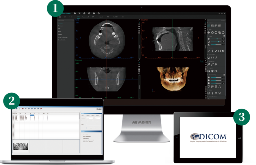
Meyer’s MyDent Viewer 3D diagnostic software implements advanced software engineering through modular design, with functional modules including multiplanar reconstruction, curved surface reconstruction, implant simulation, TMJ modelling, and 3D orthodontic simulation. Functions associated with various modules also include 3D panoramic view, 3D positioning, automatic neural tube labelling, automatic bone density measurements, automatic TMJ positioning, automatic cephalogram reconstruction, 3D airway analysis, etc.
- Easy to manage images availability of all diagnostic tools
- Simplified data managing system delivers a quick view of complex 3D data and higher efficiency of diagnosis.
- DICOM 3.0 data format output, conforms to international standards.
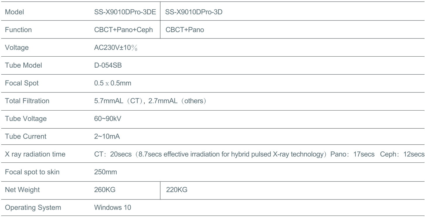
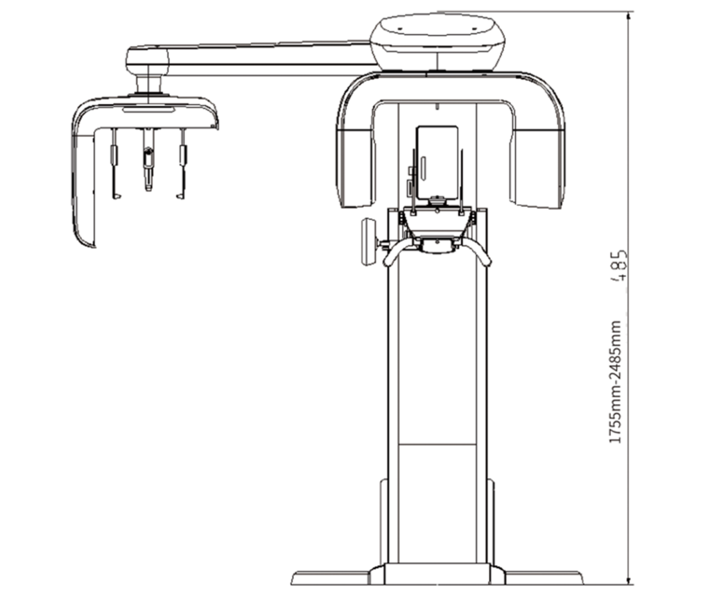
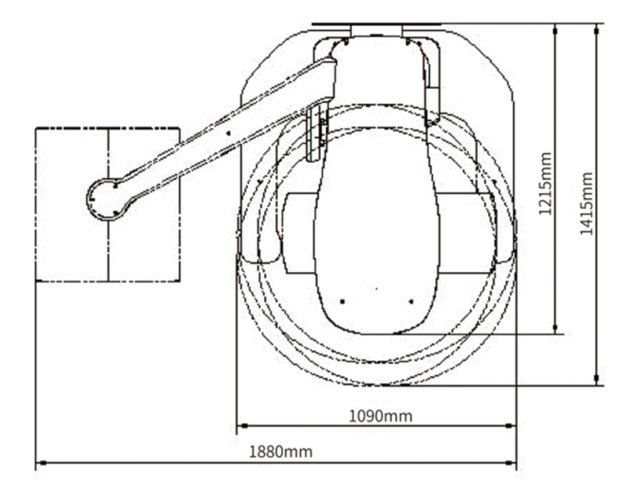
Technical specifications
